Plant Cell Microscope Labelled / 1 : As you can see in the above labeled plant cell diagram under light microscope, there are 13 parts namely. Advances in imaging plant cell dynamics plant physiology. Learn vocabulary, terms and more with flashcards, games and other study tools. As you can see in the above labeled plant cell diagram under light microscope, there are 13 parts namely, cell membrane. Parts of a microscope with functions and labeled diagram. There is also some dense.
Learn vocabulary, terms and more with flashcards, games and other study tools. Almost all plant species create their own food through the process of photosynthesis. Enjoy our articles on cell biology, the microbiology of our environment, experiments, all types of techniques and applications as well as up to date microscopy news to expand your knowledge. Cell boundaries & cell control organelles. Record the microscope images using labelled diagrams or produce digital images.
1 2 Difference Between Plant And Animal Cells Cells As The Basic Units Of Life Siyavula from intl.siyavula.com The diagram below is a plant cell as may be seen using a light microscope. Microscopy of plant cells introductory survey 1 plant cell (1): Learn about the size and function of plant and animal cells for gcse combined science, aqa. Microscopes are an essential tool to use when studying cells. Seamless pattern with animal cells. This is one of the tenets of the cell theory, a basic theory of biology. Plant cells are eukaryotic cells with a true nucleus along with specialized structures called organelles that carry out some of these differences can be clearly understood when the cells are examined under an electron microscope. The then magnified image continues up through the body tube of the microscope to the eyepiece plant and animal cells.
As you can see in the above labeled plant cell diagram under light microscope, there are 13 parts namely, cell membrane.
Here's a photo of a plant cell under an electron microscope. To examine plant cells under a microscope and find and identify different cell parts. Here's a diagram of a plant cell: Label a plant cell and match the parts with its definition. Plant cell microscope images stock photos vectors shutterstock. This is one of the tenets of the cell theory, a basic theory of biology. Microscopes are an essential tool to use when studying cells. Cell boundaries & cell control organelles. Parts of a microscope with functions and labeled diagram. However, it does not appear to be parapet wall bricks under the high power of microscope. Advances in imaging plant cell dynamics plant physiology. Answer keys are included for easy learn the structure of animal cell and plant cell under light microscope. Plant cells contain many organelles such as ribosomes, the nucleus, the plasma membrane, the cell wall, mitochondria, and chloroplasts.
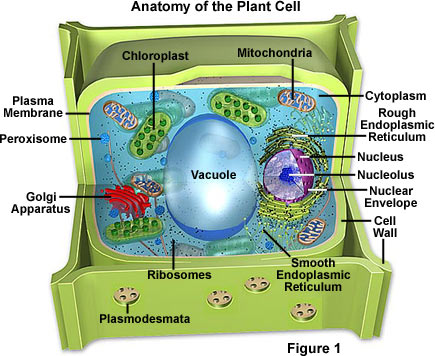
Plant cells contain many organelles such as ribosomes, the nucleus, the plasma membrane, the cell wall, mitochondria, and chloroplasts. Label a plant cell and match the parts with its definition. Examining a diagram of the plant cell will help make the differences clearer. Plant cells are very intricate structures, and they are similarities and differences between plant and animal cells. A cell is a very tiny structure which exists in living bodies.
Draw And Label A Generalized Animal Cell Shefalitayal from hi-static.z-dn.net Answer keys are included for easy learn the structure of animal cell and plant cell under light microscope. Cell is a tiny structure and functional unit of a living organism containing. All microscopes share features in common. While purchasing plant cell microscope, quality is an important parameter and this is why you need to be very cautious in looking out for a few specs such as certifications, eyepieces range, focus, stage and so on. Advances in imaging plant cell dynamics plant physiology. Vast information on microscopes, microbiology, cell biology, microscopy. Cell boundaries & cell control organelles. The diagram is very clear, and labeled the diagram is very clear, and labeled;
Plant structure and cross section botanical biology labeled diagrams collection.
But at the same time it is interpretive. Chapter 6 the cell leaving cert biology. Parts of a microscope with functions and labeled diagram. Learn vocabulary, terms and more with flashcards, games and other study tools. Magnification, however, is not the most important issue in microscopy. Advances in imaging plant cell dynamics plant physiology. Students will discover that onions are made up of cells. Your microscope has four objectives of varying magnifications (4x, 10x, 40x, and 100x) mounted on a revolving label these structures in your high power drawing. Answer keys are included for easy learn the structure of animal cell and plant cell under light microscope. Before your students use microscopes in the classroom, they should understand the names and function of each part. Microscope slide cover slip onion. Care must be taken when handling coverslips and microscope slides. Microscopy of plant cells introductory survey 1 plant cell (1):
There is also some dense. In this activity, students will create a. Continue with more related things as follows plant cell diagram without labels, microscope parts labeled and compound light. Seamless pattern with animal cells. Vast information on microscopes, microbiology, cell biology, microscopy.
Molecular Expressions Cell Biology Plant Cell Structure from micro.magnet.fsu.edu Cell boundaries & cell control organelles. Plant cell microscope image with labels cell theory plant cell. Care must be taken when handling coverslips and microscope slides. Plant cell microscope images stock photos vectors shutterstock. In truth, there are still features of plant and animal cells we're only lately. All microscopes share features in common. We have not discussed all the parts labelled since they are not part of your syllabus or will be discussed later in the syllabus. The then magnified image continues up through the body tube of the microscope to the eyepiece plant and animal cells.
Label a plant cell and match the parts with its definition.
One part of a plant cell that plays an important role in photosynthesis is a structure called a chloroplast. We have not discussed all the parts labelled since they are not part of your syllabus or will be discussed later in the syllabus. Plant cell microscope images stock photos vectors shutterstock. Cell boundaries & cell control organelles. Your microscope has four objectives of varying magnifications (4x, 10x, 40x, and 100x) mounted on a revolving label these structures in your high power drawing. Almost all plant species create their own food through the process of photosynthesis. A cell is a very tiny structure which exists in living bodies. Chapter 6 the cell leaving cert biology. Before your students use microscopes in the classroom, they should understand the names and function of each part. As you can see in the above labeled plant cell diagram under light microscope, there are 13 parts namely Observe the labeled diagram of plant cell. Care must be taken when handling coverslips and microscope slides. Plant cells contain many organelles such as ribosomes, the nucleus, the plasma membrane, the cell wall, mitochondria, and chloroplasts.
Share :
Post a Comment
for "Plant Cell Microscope Labelled / 1 : As you can see in the above labeled plant cell diagram under light microscope, there are 13 parts namely"


Post a Comment for "Plant Cell Microscope Labelled / 1 : As you can see in the above labeled plant cell diagram under light microscope, there are 13 parts namely"